FUJIFILM Europe GmbH will launch a new software version with a new function for colon polyp characterisation utilising a type of Artificial Intelligence (AI) called deep learning. FUJIFILM Corporation has already obtained a CE mark for a previous software version with the colon polyp detection function in February 2020, and both the detection and the new characterisation function have been named CAD EYE™.
The new characterisation functionality of CAD EYE together with the polyp detection function will be available with software EW10-EC02 and the compatible expansion unit EX-1 in combination with Fujifilm’s ELUXEO 7000 system and the 700 series colonoscopes. Fujifilm has already obtained a CE mark for EW10-EC02.
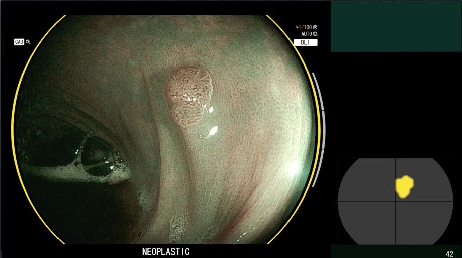
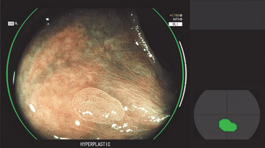
CAD EYE was developed to support real time detection of colonic polyps utilising AI technology. When a suspicious polyp is detected within the endoscopic image, a Detection Box indicates the area where the suspicious polyp has been detected accompanied by a sound signal. The new CAD EYE Characterisation will assist clinicians by generating a suggested histological prediction by displaying whether the suspicious polyp(s) in the image are hyperplastic or neoplastic.
CAD EYE functions while the moving endoscopy image is being observed and does not require complicated techniques or operations such as magnification and image capturing.
Prior to CAD EYE, Fujifilm had developed two different types of image enhancement technologies called LCI (Linked Colour Imaging) and BLI (Blue Light Imaging) for supporting detection and characterisation respectively due to their characteristics of light wavelength used. Fujifilm has applied this idea to the development of CAD EYE, and CAD EYE’s functionalities are automatically activated depending on the current observation mode.
CAD EYE Detection is activated when the clinicians are observing in White Light Mode or LCI Mode, and it automatically switches to CAD EYE Characterisation when the observation mode is changed to BLI Mode. CAD EYE can be activated or deactivated simply with just one click of the scope switch, which is also considered important when the functions are no longer necessary such as during therapeutic procedures.
The Graphical User Interface (GUI) was designed to minimise the endoscopists’ eye movements during a procedure, displaying the characterisation result and the Visual Assist Circle around the circumference of the endoscopy image. The Position Map is placed directly next to the side of the clinical image to show which area of the video image CAD EYE is focusing on.
CAD EYE Characterisation (Images below have been modified and shown for illustrative purposes only)
“Increasing the number of endoscopists who can properly detect and characterise colon polyps is one of the critical issues in the gastroenterology field,” says Prof. Helmut Neumann, Professor of Medicine and Director Interdisciplinary Endoscopy Center of University Medical Center Mainz. “With the combination of CAD EYE Detection and Characterisation, the learning curve of colonoscopy examinations can be largely improved. With the assistance of CAD EYE Detection, we can possibly see an increase in the Polyp Detection Rate (PDR), and even those who are not endoscopy experts may be brought considerably closer to this level.”
Background:
Colorectal cancer is the third most common cancer after lung cancer and breast cancer in terms of the number of cases, and the second most common cause of cancer death after lung cancer. In order to reduce the incidence of cancer, colonoscopy is widely considered the gold standard for detection of colonic neoplasia. Also, it is widely believed that accurate endoscopic diagnosis of colonic polyps could decrease the number of unnecessary polypectomies, which can lead to the reduction of medical expenses. However, there is a gap in diagnostic capability of colon polyps between endoscopy specialists and non-specialists, and filling this gap has been one of the major issues in colonoscopy.
Fujifilm is working to develop a unique collection of image processing technologies and continues to develop the practical application of AI technology, and will continue to develop and supply a wide range of products and services that meet the needs of frontline medicine in various fields, contributing to streamlining clinical work, enhancing the quality of medical care and maintaining and strengthening people's health.
